
Special Surgical Procedures II
LESSON 2: Procedures in Gynecological and Obstetrical Surgery
Section I: ANATOMY OF THE FEMALE REPRODUCTIVE SYSTEM
2-2
2-2. THE BONY PELVIS
a. The Latin word pelvis means basin. The pelvis is that part of the trunk which is surrounded by the bony pelvis. The bony pelvis (see figure 2-2) is made up of the ilium, pubis, ischium, sacrum, and coccyx. The so-called pelvic brim divides the abdominal (false) portion from the true portion of the pelvis. The abdominal (false) pelvis is the part above the arcuate line. The true pelvis is the part below this line. It forms the passageway through which the infant passes during parturition.
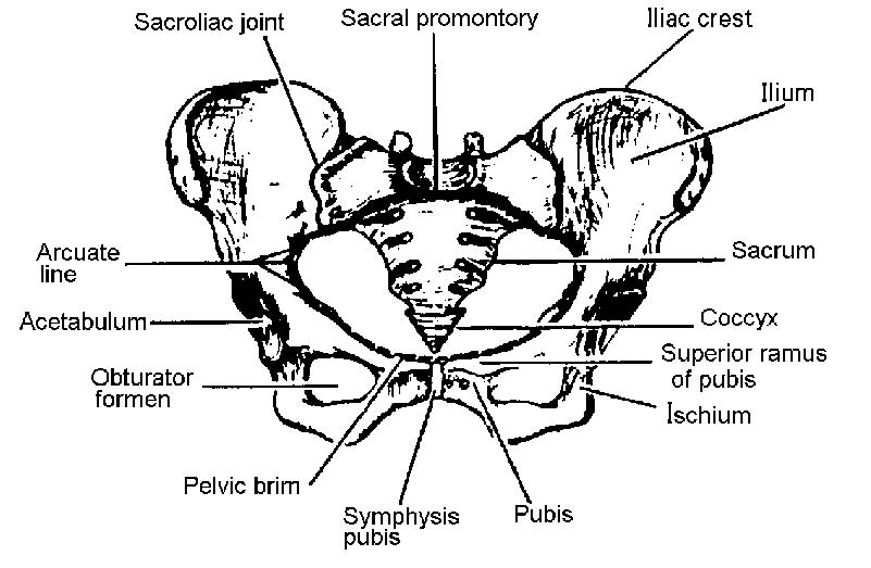
Figure 2-2. The pelvic girdle.
b. The true pelvis may be considered as having three parts: the inlet, cavity, and outlet. The muscles lining the pelvis facilitate movement of the thighs, give form to the pelvic cavity, and provide firm elastic lining to the bony pelvic framework. All organs located in the pelvis are covered by pelvic fascia. The fascia covering some muscles is dense and firm, whereas that covering other organs is thin and elastic. The nerves, blood vessels, and ureters passing through the anatomical structures are closely associated with the muscular and fascial structures.
c. The pelvic fascia may be divided into three general groups: parietal, diaphragmatic, and visceral. The parietal pelvic fascia covers the muscles of the true pelvic wall and the perineum. The diaphragmatic fascia covers both sides of the pelvic diaphragm, which is made up of the levator ani and coccygeal muscles. The visceral fascia is thin flexible fascia that covers the pelvic organs. The floor of the pelvis, known as the pelvic diaphragm, gives support to the abdominal pelvic viscera in this region. The pelvic diaphragm, consisting of the levator ani and coccygeal muscles with their respective fascial coverings, separates the pelvic cavity from the perineum. The basis of modern vaginal surgery is concerned with the function of the levator ani muscles and the provision of an effective lower outlet.
d. The levator ani muscles, varying in thickness and strength, may be divided into three parts: the iliococcygeal, the pubo-coccygeal, and the puborectal muscles. The fibers of the levator ani blend with muscle fibers of the rectum and vagina. The fibers (pubovaginal) of the pubococcygeal part of the levator ani
|
|
muscles, lying directly below the urinary bladder, are involved in the control of micturition. The pubococcygeal fibers of the levator ani control and pull the coccyx forward and assist in the closure of the pelvic outlet. The fibers pull the rectum, vagina, and bladder neck upward toward the symphysis in an effort to close the pelvic outlet and are responsible for the flexure at the anorectal junction. Relaxation of the fibers during defection permits a straightening at this junction. During parturition, the action of the levator ani directs the fetal head into the lower part of the passageway.
e. The uterus gains much of its support by its direct attachment to the vagina and by indirect attachments to nearby structures such as the rectum and pelvic diaphragm. The ligaments and muscles on each side of the uterus are the broad, round, cardinal (Mackenrodt), and uterosacral ligaments and the levator ani muscles.
