Special Surgical Procedures II

LESSON 2: Procedures in Gynecological and Obstetrical Surgery

Section I: ANATOMY OF THE FEMALE REPRODUCTIVE SYSTEM


2-4

[Previous] [Next]

Introduction


Table of Contents


Search


Exercises


Courses


About this Text


Nursing 411


Contact Us


Links

2-4. THE STRUCTURE OF THE UTERUS (FIGURE 2-3)


a. The Greek word for uterus is hystera. The uterus lies behind the bladder and in front of the rectum (see figure 2-3). The uterine body has three layers: (1) the outer peritoneal, or serous layer, which is a reflection of the pelvic peritoneum; (2) the myometrium, or muscular layer, which houses involuntary muscles, nerves, blood vessels, and lymphaticus; and (3) the endometrium, or mucosal layer, which lines the cavity of the uterus.


b. The cervix consists of a supravaginal and a vaginal portion. The supravaginal portion is closely associated with the bladder and the ureters. The vaginal portion of the cervix projects downward and backward into the top of the vaginal vault.


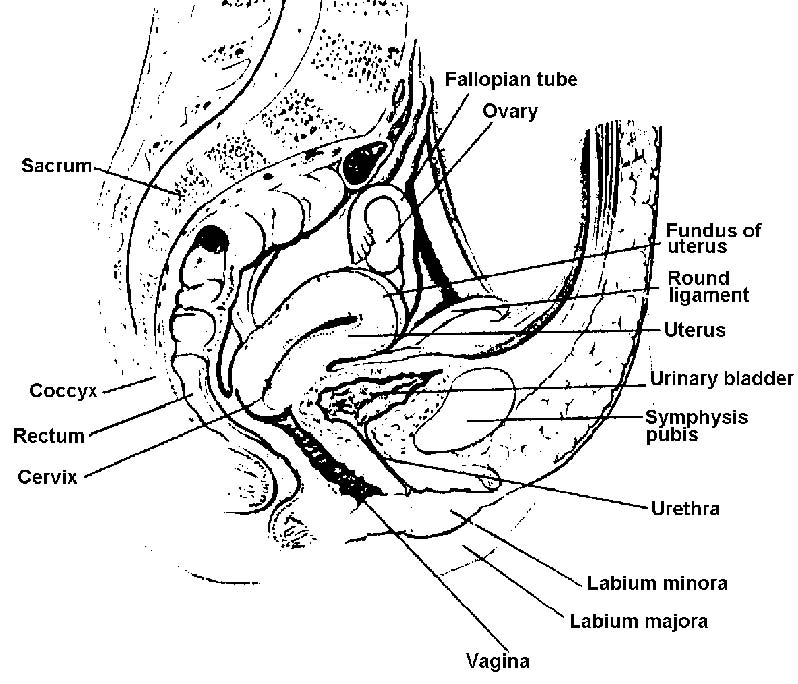
Figure 2-3. Pelvic region of female, median sagittal section.


[Previous] [Next]


These Nursing411 wings incorporate the white heart of international nursing with the
golden wings of an angel, symbolizing Nursing's selfless dedication
to the service of mankind.

The Brookside Associates Medical Education Division  develops and distributes medical information that may be useful to medical professionals and those in training to become medical professionals. This website is privately-held and not connected to any governmental agency. The views expressed here are those of the authors, and unless otherwise noted, do not necessarily reflect the views of the Brookside Associates, Ltd., or any governmental or private organizations. All writings, discussions, and publications on this website are unclassified.

© 2008 Medical Education Division, Brookside Associates, Ltd. All rights reserved

Other Brookside Products

Contact Us

Advertising on this Site