
Special Surgical Procedures II
LESSON 3: Procedures in Genitourinary Surgery
Section I: ANATOMY AND PHYSIOLOGY OF THE GENITOURINARY ORGANS
3-2
3-2. THE KIDNEYS
a. The kidneys are situated in the retroperitoneal space on the muscles of the posterior abdominal wall, one on each side of the vertebral column at the level of the twelfth thoracic to third lumbar vertebrae. Their position may vary slightly, but usually the right kidney lies lower than the left because of the space occupied by the liver. The placement of the kidneys is shown, in figure 3-1.
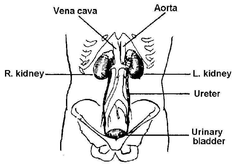
Figure 3-1. Male urinary organs in relation to other structures.
b. Each kidney is surrounded by a mass of fatty and loose areolar tissue, known as perirenal fat. Each kidney and fat capsule is surrounded by a sheath of fibrous tissue called Gerota's capsule, or renal fascia, which is connected to the fibrous tunic of the kidney by trabeculae. The kidneys are held in place by the renal fascia, which connects with the fascia of the quadratus lumborum muscle of the loins, the psoas major muscles, and the diaphragm.
c. On the medial side of each kidney there is a concave notch (called the hilum) through which the ureter, arteries, and veins enter and leave and where the renal pelvis is found.
d. The substance of the kidney (see figures 3-2 and 3-3) consists of an outer portion called the cortex, and an inner portion, called the medulla. The cortex contains the glomeruli (see figures 3-3 and 3-4) and the functioning tubules. The medulla contains many collecting tubules and papillary ducts. Each of the latter empties on a papilla within a minor calyx. Several of these join to form a major calyx. These unite to form--and therefore in turn empty into--the renal pelvis, consisting of smooth muscles lined with epithelium. The funnel-shaped renal pelvis of each kidney is continuous with the ureter below.
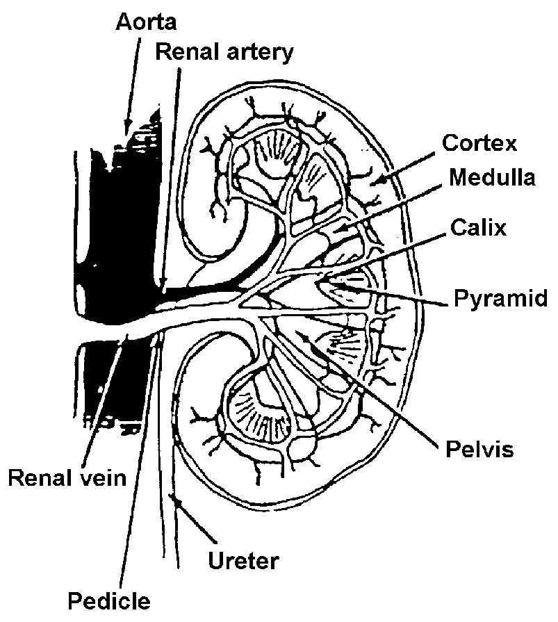
Figure 3-2. The kidney.
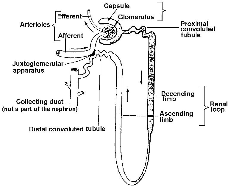
Figure 3-3. A "typical" nephron.
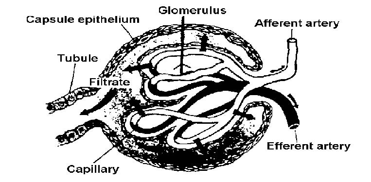
Figure 3-4. Renal corpuscle.
e. The kidneys are very vascular because one-fourth of the entire volume of blood passes through them at any one time. They receive their blood supply through the renal arteries that originate from the aorta. Each renal artery divides into several branches called afferent vessels.
f. The lymphatic supply for the most part drains into the lymph nodes that are located between the renal vessels and the aorta, and it accompanies the venous drainage.
g. The nerves of the autonomic (involuntary nervous) system carry pain sensations from the urinary organs. The nerve supply to the kidney comes from the lumbar sympathetic trunk and from the vagus nerves. Removal of the nervous pathways disrupts the ability to feel pain without impairing kidney function.
