
Special Surgical Procedures II
LESSON 1: Eye, Ear, Nose, and Throat (EENT) Surgery
Section IIi: nose SURGERY
1-28
1-28. ANATOMY AND PHYSIOLOGY OF THE NOSE
The nose is divided into the prominent external nose and the internal nose known as the nasal cavity. The chief purpose of the nose is the preparation of air for use in the lungs.
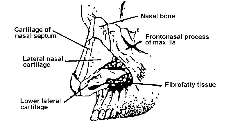
Figure 1-8. Nasal skeletal framework.
a. External Nose.
(1) The external nose projects from the face. The upper portion of the nose is formed by the nasal bones and the frontal process of the maxillae. The lower portion is formed by a group of nasal cartilages and connective tissue covered with skin. The nostrils and the tip of the nose are shaped by the major alar cartilages. The nares are separated by the columella, which is formed by the lower margin of the septal cartilage, the medial parts of the major alar cartilages and the anterior nasal spine, all of which are covered by skin.
(2) The nasal septum is composed of three structures: the nasal cartilage, the vomer bone, and the perpendicular plate of the ethmoid bone. The septum is covered by mucous membrane on either side. The deviated or fractured septum may be repaired surgically by mobilization of the fracture or removal of the deformed cartilage or bone.
b. Internal Nose.
(1) The internal nose or nasal cavity is divided into two parts at its midline by the nasal septum. The nasal cavity communicates with the outside by its external openings, called the anterior nares. The nares open into the nasopharynx behind through the choanae. The nasal cavity is also associated with each ear by means of the eustachian tube and with the paranasal air sinuses (see figure 1-9) (frontal, maxillary, ethmoid, and sphenoid) via their respective orifices (meatuses). The nasal cavity communicates with the conjunctive through the nasolacrimal duct as well.
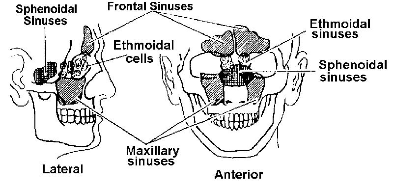
Figure 1-9. The paranasal sinuses (lateral and anterior aspects).
(2) The nasal cavity is separated from the lingual cavity by the hard and soft palates and from the cranial cavity by the ethmoid bone. The nasal cavity is held together by periosteal covering and by perichondrium, which extends over the cartilages.
c. Turbinate Bones and Sinuses.
(1) The turbinate bones of the nasal structure are arranged one above the other, separated by grooves (the meatuses). These act as drainage passages of the accessory sinuses and are
known as the sphenoethmoidal recess and the superior, middle, and inferior meatus, respectively.
(2) The nasal sinuses serve as air spaces and communicate with the nasal cavity via the meatuses. Anteriorly, on each side of the skull, the frontal sinus, the anterior ethmoid cells, and the maxillary sinus (antrum of Highmore) drain into the middle meatus; posteriorly, the ethmoid cells and the sphenoid sinus drain into the superior meatus and the sphenoethmoidal recess. A passageway for the flow of air is provided by the irregular air spaces present between these structures. Because of their shape, the air is forced to flow in thin airwaves.
d. Nerve and Blood Supplies.
(1) The sensory nerve supply of the nasal cavity is derived from the trigeminal nerve.
(2) The nose and sinuses receive their blood supply from branches of the internal maxillary artery. There are masses of communicating veins below the epithelial layer of the turbinated bones, and those veins lying just beneath the mucosa anastomose (communicate) freely. Dilatation of the superficial veins may cause the turbinated bone mucosa to swell, whereas contraction of these vessels may cause the mucosa to shrink.
