
Special Surgical Procedures II
LESSON 1: Eye, Ear, Nose, and Throat (EENT) Surgery
Section II: EAR SURGERY
1-18
Section II. EAR SURGERY
1-18. GENERAL ANATOMY AND PHYSIOLOGY OF THE EAR
The ear (see figure 1-5) is made up of three distinct divisions: the external ear, the middle ear, and the inner ear. The middle and inner ear structures are situated in the temporal bone cavity.
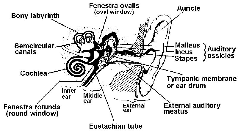
Figure 1-5. Parts of the ear
a. External Ear. The external ear consists of an auricle, or pinna, and an external auditory meatus (a tube ending at the tympanic membrane or ear drum). The auricle collects the sound vibrations in the air and sends them through the external canal to the ear drum.
b. Tympanic Membrane. The tympanic membrane (see figure 1-6) is a tri-layered membrane stretched across the end of the external meatus. The sound waves come through the meatus and vibrate the membrane.
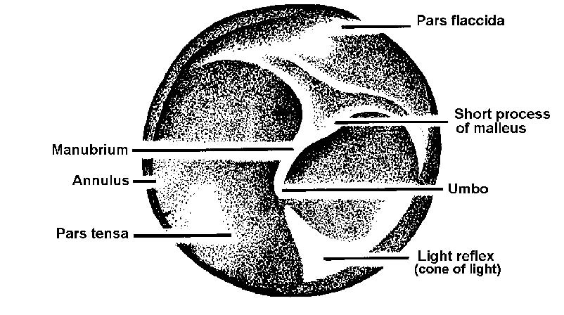
Figure 1-6. Landmarks of right tympanic membrane.
c. Middle Ear. Inside the tympanic membrane is a narrow, irregular, oblong, air-conditioned cavity in the tympanic part of the temporal bone. This air-filled space contains three small bones, which transfer the vibrations from the tympanic membrane to the inner ear. Figure 1-7 shows the ossicles of the middle ear.
d. Inner Ear. The inner ear is a complex structure located in the petrous portion of the temporal bone. It is made up of two distinct parts, each of which contains its own kind of fluid. Sound vibrations carried by the bones of the inner ear are transferred by way of the oval window to the fluid in the cochlea and received through a fine membrane by the organ of Corti, the delicate neural end organ for sound. A second function of the inner ear is the maintenance of balance, controlled by the movement of fluid in the labyrinth in relation to neuroepithelial cells.
e. Temporal Bone. The temporal bone houses the middle and inner ear as well as the mastoid sinuses.
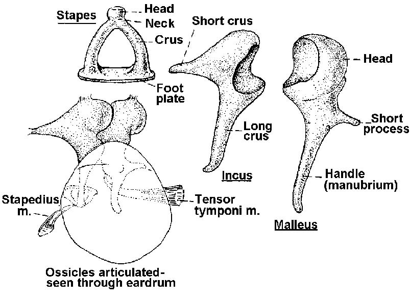
Figure 1-7. Ossicles of the middle ear (separated and articulated).
