
Special Surgical Procedures II
LESSON 1: Eye, Ear, Nose, and Throat (EENT) Surgery
Section I: EYE SURGERY
1-1
Section I. EYE SURGERY
1-1. INTRODUCTION
a. General. The anatomy, physiology, and the location of the eye make surgery upon the eye a highly specialized field of surgery. Therefore, procedures done by the specialist when assisting with eye surgery differ from procedures used for other surgical specialties. However, the principles of asepsis and safe, skillful care apply as in all other surgery. The ensuing text presents a discussion of the necessary considerations that are applicable in the majority of cases in this specialty.
b. Special Care of Instruments. The specialist is to use exacting care when working with instruments for eye, ear, nose, and throat surgery because most of these instruments are delicate. Sharp surfaces of these instruments must be preserved to ensure the success of the operative procedure. The specialist is to follow local policy in the care and handling of these instruments.
c. Anatomy and Physiology of the Eye. The eye is also referred to as the eyeball or globe. In the adult, it is slightly less than one inch in its longest diameter. See figure 1-1 for parts of the eye.
(1) The lids and anterior surface of the eye, except for the center, are covered by the conjunctiva.
(2) The cornea forms the anterior center of the eye and transmits and refracts light. Behind it, the anterior chamber contains the iris (which gives eye color and forms the pupil) and the aqueous humor.
(3) The lens focuses light on the retina allowing for near and far vision.
(4) The posterior chamber contains the jelly-like vitreous humor, which helps give rigidity to the eye.
(5) The retina receives light and converts it to impulses to the brain via the optic nerve.
(6) The main body of the eye is made of three layers called tunics. The external tunic includes the sclera (the white part of the eye) and clear cornea. The middle tunic includes the choroid, the ciliary body, and the iris. The iris is the colored part that changes the aperture size over the eye lens. The internal tunic is sometimes called the nervous covering, but is usually referred to as the retina. The retina is a thin network of nerve cells and fibers that receives the images of objects the eye is seeing.
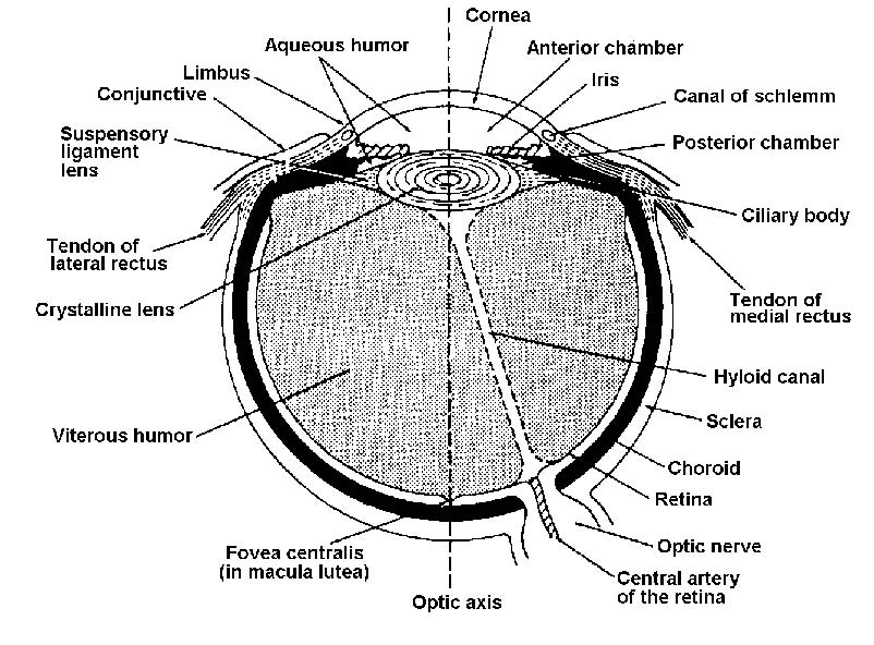
Figure 1-1. Parts of the eye.
