
Integumentary System
Lesson 1: Anatomy and Physiology of the Integumentary System
1-4
1-4. FUNCTIONS OF THE SKIN
a. Sensation.
(1) Sensation refers to a state of awareness of conditions of the body. Four prerequisite conditions must be present in order for a sensation to occur:
(a) Stimulus (or change in environment)--something capable of initiating a response by the nervous system.
(b) Receptor or sense organ--something must be able to pick up the stimulus and convert the stimulus to a nerve impulse. A sense receptor or sense organ is specialized tissue that is very sensitive to conditions affecting the body.
(c) Conductor--the impulse must be conducted along a nerve pathway from the receptor or sense organ to the brain.
(d) Translator--a region of the brain must translate the impulse into a sensation.
(2) Receptors can be classified according to their location. Exteroceptors are near the surface of the body. Viscereceptors or enteroceptors are located in the blood vessels and viscera. Proprioceptors are receptors located in muscles, tendons, joints, and the external ear. Exteroceptors, located near the surface of the body, provide information about the external environment. These receptors, sensitive to stimuli outside the body, transmit sensations of hearing, sight, smell, taste, touch, pressure, temperature, and pain. The exteroceptors located in the skin provide the sensations of pain, touch, temperature, and pressure.
(a) Pain. Receptors for pain are not only found in the skin but in practically every tissue of the body. These receptors may be stimulated by stimuli for other sensations. When the receptors for touch, pressure, heat, and cold reach a certain threshold, they stimulate the sensation of pain also. Since pain receptors are sensitive to all stimuli, these receptors perform a protective function by telling the body of changes that may be a danger to the body. There are two types of pain receptors: somatic pain receptors and visceral pain receptors. Somatic pain comes from the stimulus of receptors in the skin and receptors in skeletal muscles, joints, tendons, and fascia. Visceral pain comes from stimulation of receptors in the viscera.
(b) Touch. Touch sensations generally result from stimulation of tactile receptors in the skin or the tissues immediately beneath the skin. Light touch refers to the ability to recognize exactly what point of the body is touched. Crude touch refers to the ability to perceive that something has touched the skin although its exact location, shape, size, or texture cannot be determined. Receptors for touch include root hair plexuses, free nerve endings, Merkel's discs, Meissner's corpuscles, and end organs of Ruffini. Root hair plexuses are dendrites arranged in networks around the roots of hairs. Free nerve endings are found everywhere in the skin. Merkel's discs are disc-like formations of dendrites attached to deeper layers of epidermal cells. Meissner's corpuscles, located in the dermal papillae of the skin, are egg-shaped receptors containing a mass of dendrites enclosed by connective tissue.
(c) Temperature. These receptors are sensitive to heat and cold. It is thought that temperature receptors are free nerve endings.
(d) Pressure. Pressure receptors generally come from the stimulation of touch receptors in deeper tissues. Sensation from these receptors lasts longer and is spread over a greater area than the sensation from touch receptors.
b. Protection. The function of skin is to protect the body's underlying structures from bacterial invasion, drying out, and harmful light rays. The acid mantle (pH 4.2 to 5.6) on the skin surface protects the body from bacteria and irritants. Skin keeps the body from excessive water and electrolyte loss.
c. Thermoregulation. Man is capable of maintaining a relatively constant body temperature (37°C or 98.6°F). If man is in an environment of 100°F, sensing devices in the skin called receptors pick up the heat stimulus and send a message to the brain. A temperature-regulating area of the brain sends nerve impulses to the sudoriferous glands that cause these glands to produce more perspiration. As the perspiration evaporates from the skin surface, the skin surface is cooled, and the body temperature is maintained.
d. Types of Tissues. See figure 1-5.
(1) General. Tissue can be defined as a group of similar cells and their intercellular substance functioning together to perform a specialized activity. Some tissues move body parts. Other tissues move food through body organs while some tissues protect and support the body. Other tissues produce chemicals such as enzymes and hormones. Body tissues are classified by function and structure into four principal types: epithelial tissue, connective tissue, muscular tissue, and nervous tissue. Two of these types will be examined in this subcourse: epithelial tissue, which covers body surfaces or tissues, lines body cavities, and forms glands; and connective tissue, which protects and supports the body and its organs and binds organs together.
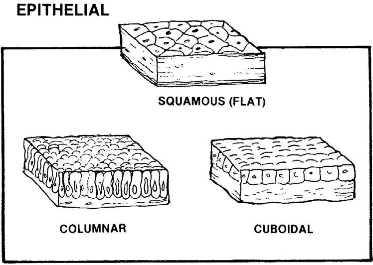
Figure 1-5. Types of skin tissues.
(2) Epithelial tissues. The tissues on the outer surfaces of the body are faced with epithelial cells. The deep surfaces of the skin are layered with connective tissue which strengthens membranes. There are two types of epithelium tissue: mucous membrane epithelial tissues (covering and lining epithelium) and serous membrane epithelial tissues (glandular epithelium). Both types of epithelial tissue consist of tightly packed cells with very little intercellular material between the cells. The cells are arranged in continuous sheets with either one layer or several layers. It is possible for nerves to extend through the sheets but blood vessels do not. The underlying connective tissue holds the epithelial tissue in place and prevents it from being torn. A thin extra cellular cellar called the basement membrane glues the epithelium and the connective tissue together. All epithelial cells face a certain amount of wear, tear, and injury. To replace themselves, epithelial cells divide and produce new cells.
(a) Mucous membrane epithelial tissues. These epithelial cells line the tubes and cavities that open to the exterior of the body; for example, the mouth, nose, intestinal tract and urinary and reproductive tracts are lined with mucous membrane epithelial cells. Simple epithelial cells (tissue cells arranged in a single layer) line the air sacs of the lungs where oxygen is exchanged with carbon dioxide. This type of lining is present in the part of the kidney that filters the blood. The inner surfaces of interior parts of the ear are lined with epithelial cells. This lining, as evidenced by these examples, is found in body parts that have very little wear and tear. Another function of mucous membrane epithelial cells is to secrete mucus that serves as protection against the entry of foreign particles into the body. Some epithelial cells are ciliated. Cells with hair-like processes called cilia are found in some parts of the respiratory tract. These cilia wave in unison and move mucus plus trapped foreign particles toward the throat where the substance can either be swallowed or coughed out. This is the process of filtering air before it enters the lungs.
(b) Serous membrane epithelial tissues. These tissues are better known as glands or glandular epithelium. Serous membrane epithelial cells may be one cell or a group of specialized epithelial cells whose function is to secrete substances
into ducts, onto a surface, or into the blood. Glandular cells work to produce substances and expend energy in that effort. Glands that secrete substances into ducts (tubes) that empty at the surface of covering and lining epithelium or directly onto a surface are classified as exocrine glands. Products secreted by exocrine glands include mucus, perspiration, oil, wax, and digestive enzymes. Those glands that have no ducts and secrete their substances directly into the blood are classified as endocrine glands. Endocrine glands secrete hormones. Examples of endocrine glands include the pituitary, thyroid, and adrenal glands. Serous membrane epithelial tissues also cover some organs of the body: the pleura enclose the lungs; the pericardium covers the heart; and the peritoneum lines the abdominal cavity. These membranes secrete a thin fluid that prevents friction when organs are in contact with one another.
(3) Connective tissues. Connective tissue (figure 1-6) primarily binds and supports, is highly vascular, and has a rich blood supply. An exception is cartilage that is avascular (no blood vessels). Cells in connective tissue are widely scattered rather than closely packed, and there is a lot of intercellular material. The general functions of connective tissue are protection, support, and the binding together of various organs. These tissues anchor and support organs and cover bone and cartilage. Two types of connective tissue will be explored in this lesson: skeletal connective tissue and fascial (fibrous) connective tissue.
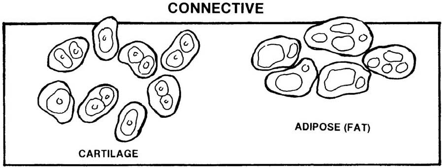
Figure 1-6. Connective tissue.
(a) Skeletal connective tissue. Synovial membranes line the joint cavities. Like serous membranes, these membranes do not open to the exterior. They cover tendons and secrete a thin lubricant fluid. Synovial membranes are composed of loose connective tissue with elastic fibers and varying amounts of fat. Synovial fluid is secreted by synovial membranes. This fluid lubricates the ends of bones as they move at joints and nourishes the articular cartilage covering the bones that form the joints. Periosteum is a connective membrane classified as synovial connective membrane. Periosteum covers bone, and perichondrium covers cartilage.
(b) Fascial (fibrous) connective tissue. The term fascia refers to a sheet or a broad band of fibrous connective tissue that is under the skin or around muscles and other organs of the body. There are three types of fascia: superficial fascia or subcutaneous layer (immediately deep in the skin); deep fascia (the most extensive of the three types); and subserous (visceral) fascia (located between the internal layer of deep fascia and a serous membrane).
1 Superficial fascia (subcutaneous layer). This type of connective tissue covers the entire body and varies in thickness in different regions. It is quite thin on the back of the hand but quite thick on the abdominal wall. The functions of this tissue include serving as a storehouse for water and for fat; forming an insulating layer to protect the body from loss of heat; providing a layer of protection from blows to the body; and providing a pathway for nerves and vessels.
2 Deep fascia. This type of connective tissue lines the body wall and extremities and holds muscles together, separating them into functioning groups. Deep fascia allows free movement of muscles, carries nerves and blood vessels, fills spaces between muscles, and sometimes provides the origin for muscles.
3 Subserous (visceral) fascia. This type of tissue forms the fibrous layer of serous membranes, covering and supporting the viscera, and attaching the parietal layer of serous membranes to the internal surface body wall.
