
1-2
1-2. THE HEART
The heart, designed to be a highly efficient pump, is a four-chambered muscular organ, lying within the chest, with about 2/3 of its mass to the left of the midline. It lies in the pericardial space in the thoracic cavity between the two lungs. In size and shape, it resembles a man's closed fist. Its lower point, the apex, lies just above the left diaphragm. Refer to Figure 1-2 as you continue to read.
a. Heart Layers. The pericardium is a double walled sac enclosing the heart. The outer fibrous surface gives support, and the inner lining prevents friction as the heart moves within its protecting jacket. The lining surfaces of the pericardial sac produce a small amount of pericardial fluid needed for lubrication to facilitate the normal movements of the heart.
b. Heart Wall. The walls of the heart is composed of three distinct layers an outer epicardium- which corresponds to the visceral pericardium it protects the heart by reducing friction, a middle layer the myocardium consist mostly of cardiac muscle tissue that pumps blood out of the heart chambers, an inner layer endocardium consist of epithelium and connective tissue that contains many elastic and collagenous fibers.
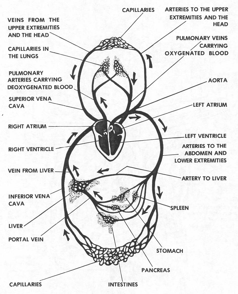
Figure 1-1. The circulatory system.
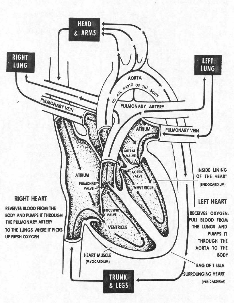
Figure 1-2. The heart.
c. Heart Chambers. There are four chambers in the heart. These chambers are essentially the same size. The upper chambers, called the atria, are seemingly smaller than the lower chambers, the ventricles. The apparent difference in total size is due to the thickness of the myocardial layer. The right atrium communicates with the right ventricle; the left atrium communicates with the left ventricle. The septum (partition), dividing the interior of the heart into right and left sides, prevents direct communication of blood flow from right to left chambers
or left to right chambers. This is important, because the right side of the heart receives un-oxygenated blood returning from the systemic (body) circulation. The left side of the heart receives oxygenated blood returning from the pulmonary (lung) circulation. The special structure of the heart keeps the blood flowing in its proper direction to and from the heart chambers.
d. Heart Valves. The four chambers of the heart are lined with endocardium. At each opening from the chambers this lining folds on itself and extends into the opening to form valves. These valves allow the blood to pass from a chamber but prevent its return. The atrioventricular valves, between the upper and lower chambers, are within the heart itself. The semilunar valves are within arteries arising from the right and left ventricles.
(1) Atrioventricular valves. The tricuspid valve is located between the right atrium and right ventricle. It has three flaps or cusps. The bicuspid valve or mitral valve is located between the left atrium and left ventricle. It has two flaps or cusps.
(2) Semilunar valves. The pulmonary semilunar (half-moon shaped) valve is located at the opening into the pulmonary artery that arises from the right ventricle. The aortic semilunar valve is located at the opening into the aorta that arises from the left ventricle.
